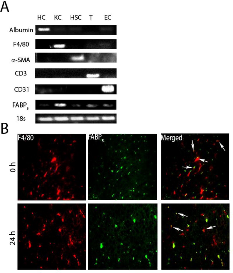
Figure 1. Cell purity and co-localization of FABP5 expression with expression of the hepatic macrophage marker F4/80.

A: Cell colonies were obtained from the livers of C57Bl/6 mice as described in materials in methods. The mRNAs of hepatocyte (HC) marker albumin, hepatic Kupffer cell (KC) marker F4/80, hepatic stellate cell (HSC) marker α -SMA, T-cell (T) marker CD3, endothelial cell (EC) marker CD31, and FABP5 were detected by reverse transcription polymerase chain reaction. B: Immunohistochemistry was performed on sections of liver from C57Bl/6 (WT) mice treated with saline as control (0 h) or LPS for 24 hours. Antibodies to the hepatic macrophage marker F4/80 and FABP5 were used. Merged image shows colocalization of F4/80 and FABP5 expression. Micrographs are at 40x magnification. Arrows point to hepatic macrophages.
