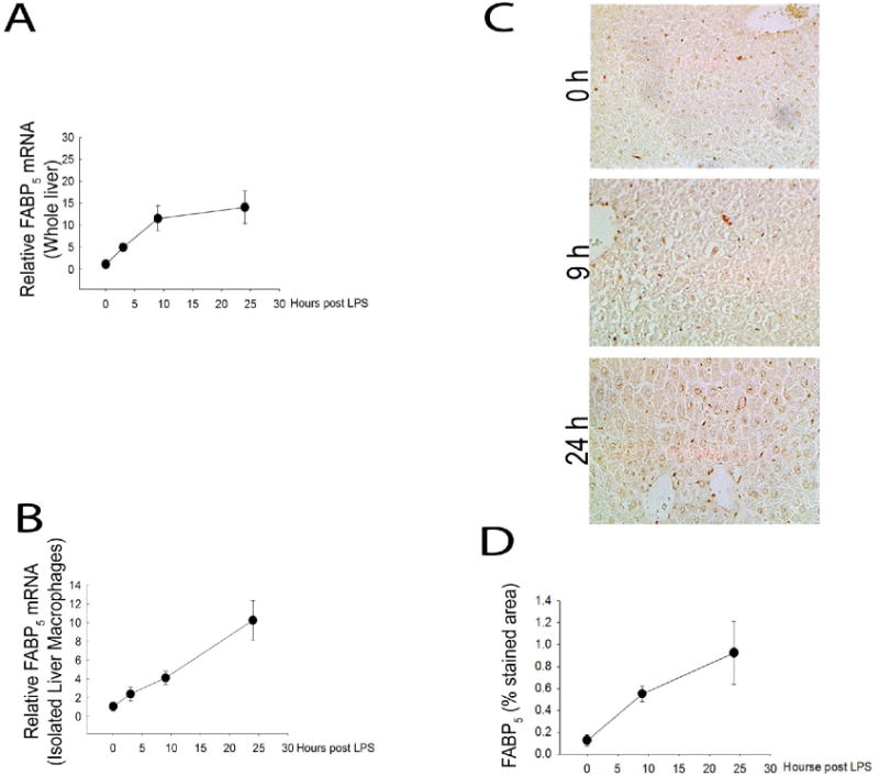
Figure 2. LPS promotes hepatic FABP5 expression.

Wildtype and FABP5−/− mice were treated with saline (control) or LPS for 3, 9 or 24 hours. A: Control whole liver FABP5 mRNA expression was analyzed via quantitative PCR and normalized to the housekeeping gene 18S rRNA. B: WT liver macrophages were isolated as stated earlier and FABP5 mRNA expression was analyzed via quantitative qPCR and normalized to 18s rRNA. C: Immunohistochemistry was performed on sections of liver tissue from WT mice treated with LPS for 0, 9, or 24 hours using an antibody to FABP5. D: Percent stained area was determined by image J analysis in three separate measurements from each time point.
