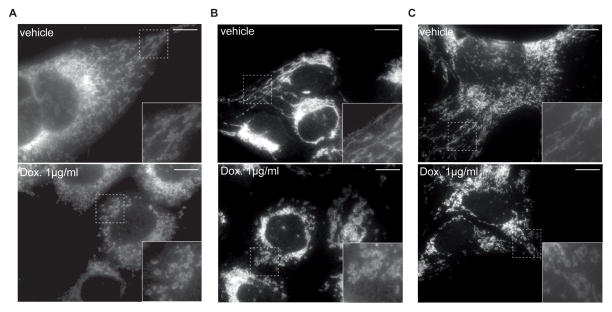
Figure 2. Impact of doxycycline on mitochondrial morphology in cultured cells.
Mitochondria in Hepa 1-6 (A), HeLa (B), and GT1-7 (C) cells appear more fragmented after treatment with doxycycline at the indicated concentration.
Mitochondria were stained with TOM40 antibody. Insets showed higher magnification of the image, marked by the rectangle. Scale bar represents 10μM.

