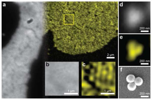
Figure 2.

(a) Comparison between direct 2P (gray-scale) and 2P-SPIM (pseudo-color) images of same fluorescence nanosphere phantom. A region within the sample as highlighted by the yellow box was magnified to compare (b) direct 2P microscopy and (c) 2P-SPIM. The monolayer of the 200 nm fluorescent spheres can be resolved using 2P-SPIM. Smaller clusters of spheres were also imaged using (d) direct 2P microscopy and (e) 2P-SPIM to compare structures visualized by (f) SEM microscopy.
