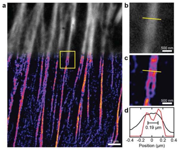
Figure 3.

(a) Direct 2P microscopy (grayscale) and 2P-SPIM (pseudo-color) images of the actin cytoskeleton in the same HeLa cell stained with Alexafluor 546 phalloidin. (b) Magnified direct 2P microscopy image of the region within the yellow box. (c) Magnified 2P-SPIM microscopy image of the region within the yellow box. (d) One-dimensional profiles of imaged microtubules at the location highlighted by the yellow lines. Resolution is 180 nm in 2P-SPIM image.
