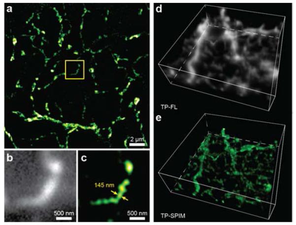
Figure 4.

Volumetric imaging using 2P-SPIM. (a) Two-dimensional 2P-SPIM reconstructed image of ganglion cell dendrites in ground squirrel retina at a depth of 100 μm from the vitreal surface. A magnified comparison (within the yellow box) of (b) direct 2P microscopy (grayscale) and (c) 2P-SPIM (pseudo-colored) images. The 2P-SPIM reconstruction can resolve the object with a size of ~145 nm, which is still much beyond its diffraction limit, at the depth of 100 μm. Three-dimensional image stacks at depths from 94 μm to 110 μm in the retinal tissue were created using both (d) direct 2P microscopy (grayscale) and (e) 2P-SPIM (pseudo-colored). A total of 16 slices acquired at 1 μm depth interval were stacked to form the volumetric images. The cell soma, at top, is removed. Z-axis resolution is limited by the 2P point spread function.
