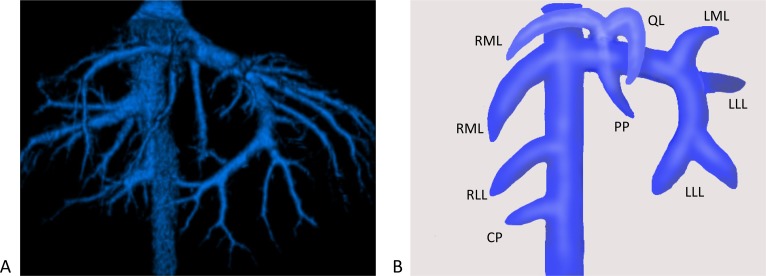
Fig. 3.
(A) Ventral-dorsal hepatic vein of a normal dog. (B) Schema of the hepatic vein, with a main branch of the hepatic vein to each lobe shown. PP, papillary process of the caudate lobe; LLL, left lateral lobe; LML, left medial lobe; QL, quadrate lobe; RML, right medial lobe; RLL, right lateral lobe; CP, caudate process of the caudate lobe.

