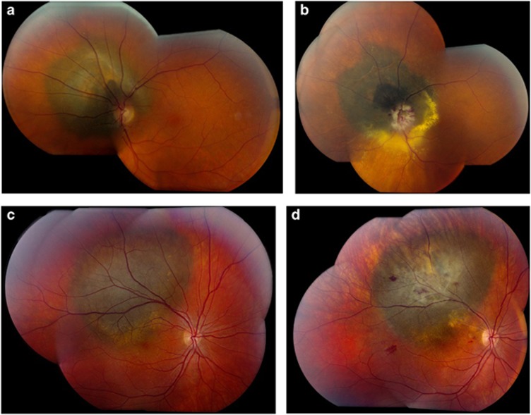
Figure 1.
(a): Fundus photograph of a 64 year-old female patient with a melanoma encircling 8 clock hours of the left optic disc before stereotactic radiosurgery. Visual acuity at presentation was 6/12. (b): Fundus photograph of the patient in figure 1a, 2 years post-stereotactic radiosurgery showing the melanoma to be in regression with oedema and haemorrhage involving the optic disc consistent with radiation optic neuropathy. Final visual acuity was counting fingers. (c): Fundus photograph of the right eye of a 28 year-old male patient with a juxtafoveal melanoma before proton beam therapy. Visual acuity at presentation was 6/12. (d): Fundus photograph of the patient in figure 1c, 2 years following proton beam therapy showing the melanoma to be in regression with scattered retinal haemorrhages and a small vascular occlusion consistent with radiation retinopathy. Visual acuity at this stage was 6/18.

