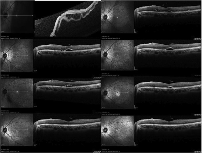
Figure 3.
Serial fundus infrared reflectance (IR) and spectral-domain optical coherence (SDOCT) tomography images showing the natural course of residual SRF blebs after scleral buckling surgery for macula-off rhegmatogenous retinal detachment (RRD). (Top left) Preoperative IR and SDOCT images of a 37-year-old patient with macula-off RRD. Undulation of the photoreceptor layer and severe cystoid changes in the outer nuclear layer were present. Visual acuity was counting fingers. (Top right) At 1 month postoperatively, SRF blebs were observed. Visual acuity was 0.32 logMAR. (Second row left) Three months after surgery, visual acuity was 0.32 logMAR. (Second row right) At 6 months, visual acuity was 0.24 logMAR. (Third row left) At 9 months, visual acuity was 0.18 logMAR. Note the progressive elongation of the photoreceptors' outer segments. (Third row right) One year after surgery, visual acuity was 0.18 logMAR. Note the bleb width decrease. (Bottom left) At 18 months, visual acuity was 0.08 logMAR. The SRF bleb finally regressed leaving focal defects in the photoreceptors' inner segment/outer segment (IS/OS) junction line. (Bottom right) Two years after surgery, visual acuity was 0.08 logMAR. Note the apparent complete recovery of the IS/OS junction band.

