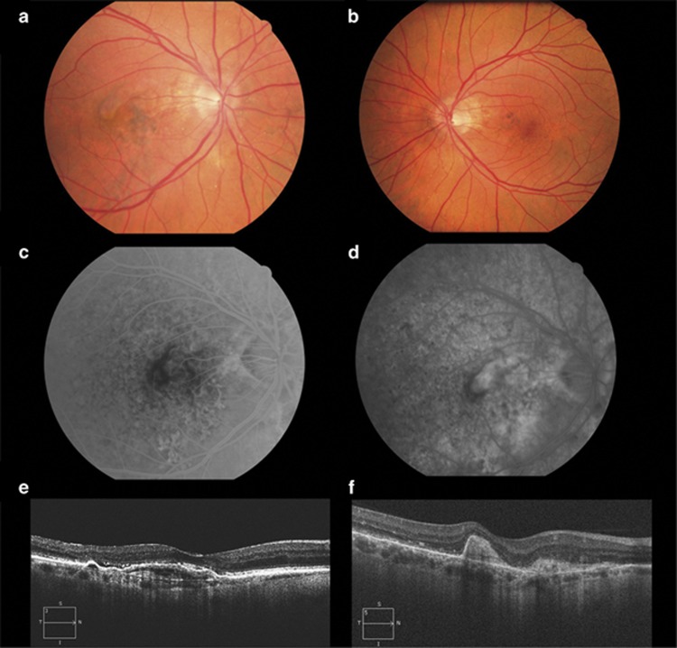
Figure 2.
Colour fundus photographs show bilateral angioid streaks, OD (a) and OS (b) and a justafoveal choroidal neovascular membrane in OD (a). Fundus fluorescein angiography shows an early well-demarcated lesion (c) with late leakage (d) corresponding to a predominantly classic choroidal neovascularisation. Spectral-domain optical coherence tomography reveals a subfoveal pigment epithelium detachment with associated subretinal fluid (e) at baseline and resolution of the fluid and subretinal fibrosis after treatment (f). Diagnostic work-up to rule-out systemic associations was negative.

