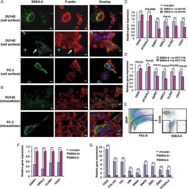
Fig. 4.
Characterization of SSEA-4 expressing cells. (A and B) Localization of SSEA-4 on DU145 and PC3 cells. (A) Adherent DU145 and PC3 cells were surface labeled with SSEA-4, fixed and then permeabilized, followed by staining with Alexa Fluor 488-labeled secondary antibody and Rhodamine-labeled phalloidin. Cells were mounted in DAPI containing mounting media as described in Methods. Note the conversion of cells from a colonial epithelial morphology (less than symbol) into a polarized, migratory mesenchymal morphology (up-arrow) and the localization of SSEA-4 to the plasma membrane of polarized cells. (B) Adherent cells were fixed, permeabilized, labeled with IPS-K-4A2B8 followed by staining with Alexa Fluor 488-labeled secondary anti-mouse IgM antibody and Rhodamine-labeled phalloidin. Note weak staining of SSEA-4 in the cytoplasm of all DU145 cells. In contrast to DU145 cells, most PC3 cells that were negative for cell surface SSEA-4 did not express cytoplasmic SSEA-4. Scale bars: 10 µm. (C and D) Expression of several EMT-related genes are modulated in SSEA-4+ and SSEA-4− DU145 as well as HCT-116 cells. Relative gene expression of EMT markers on SSEA-4+ and SSEA-4− (C) DU145 as well as (D) HCT-116 cells shows higher expression of mesenchymal marker (CDH2) in SSEA-4+ subset and epithelial markers (ESRP1, GRHL2, CLDN7 and CDH1) in SSEA-4− cells. Also note the increase in the expression of ST3GAL2 in SSEA-4+ cells. Shown are data from one representative biological experiment performed in technical replicates, quantified relatively to SSEA-4− cells and normalized to GAPDH. Error bars denote mean ± standard error. p-values were calculated using Student's t-test. (E) Display of SSEA-4 expression on primary prostate cancer cells. Sort windows were set and cells were sorted on a FACS Aria cell sorter. (F and G) Differential expression of EMT-related genes in SSEA-4+ and SSEA-4− primary prostate tumor cells. (F) Relative gene expression of EMT-related markers in SSEA-4+ and SSEA-4− cells shows higher expression of epithelial markers in SSEA-4− cells. (G) In contrast, SSEA-4+ cells show an increased expression of mesenchymal markers (CDH2, COL1A2, FN1 and VIM), EMT markers (SNAI1, SNAI2, ZEB1 and ZEB2) and SSEA-4 synthesizing enzyme ST3GAL2. Data are shown from one patient sample performed in technical replicates, quantified relatively to SSEA-4− cells and normalized to GAPDH. Error bars denote mean ± standard error. p-values were calculated using Student's t-test.

