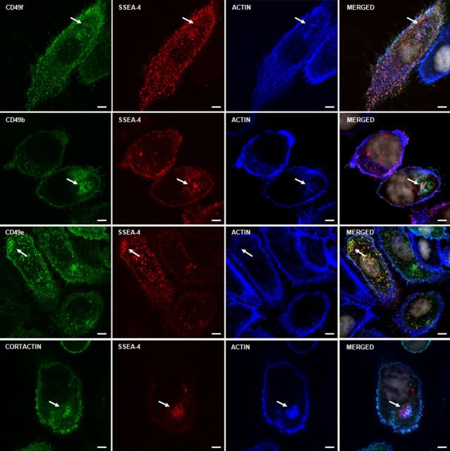
Fig. 5.
Localization of SSEA-4, F-actin and integrins on PC3 cells. Three color staining of adherent PC3 cells with anti-integrins, phalloidin (F-actin) and mAb IPS-K-4A2B8 shows that integrins CD49f, CD49e and CD49b colocalize with SSEA-4 and F-actin accumulating spots. Arrows point to accumulation of integrins at F-actin and SSEA-4 accumulating structures. Scale bars: 5 µm.

