
Fig. 5.
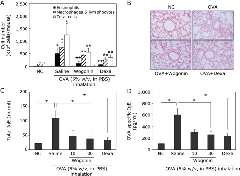
Effects of wogonin on OVA-induced airway inflammation. (A) BAL fluids were collected 48 h after the last OVA challenge of mice. Cells were isolated by cyto-spinning and stained with Diff-Quick. Cell numbers were assessed by counting cells directly under a light microscope in at least five squares of a hemocytometer, after excluding dead cells staining with trypan blue. NC, normal control mice; OVA, OVA-sensitized and challenged mice, Wogonin, OVA-sensitized and challenged mice treated with wogonin (30 mg/kg); Dexa, OVA-sensitized and challenged mice treated with dexamethasone (4 mg/kg). The values are expressed as the means ± SEM (n = 6 per group). *Significant difference from NC, p<0.05, and **significant difference from OVA p<0.05. (B) Histological examination of lung tissue was performed 48 h after the last OVA challenge. Lung tissues were fixed and stained with H&E solution. (C, D) Samples of serum were collected 48 h after the last OVA challenge. Levels of total IgE (C) and OVA-specific IgE (D) in serum were analyzed by ELISA. *p<0.05.