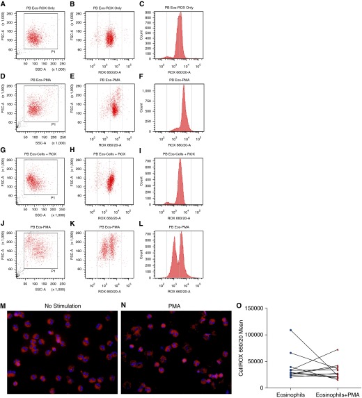
Figure 2.
Intracellular reactive oxygen species (ROS) in blood eosinophils. Flow cytometry analysis of eosinophils stained with CellROX showed change in cell size and granularity with PMA. (A–C) Data obtained from the eosinophils from one subject. (G–H) Data obtained from another subject. In both cases, eosinophils were stained with CellROX without stimulation. (D–F and J–L) The corresponding data in PMA-treated and CellROX-stained eosinophils of these two individuals (n = 12). (E, F, K, and L) Data from two subjects with different degrees of segregation of two populations of eosinophils with respect to their ROS content after PMA stimulation. (M and N) Fluorescent micrographs of unstimulated and PMA-stimulated eosinophils, respectively, stained with CellROX and 4′,6-diamidino-2-phenylindole. (O) There was no change in the intracellular ROS in PMA-induced eosinophils (n = 12). FSC-A, forward scatter; PB Eos, peripheral blood eosinophils; PMA, phorbol myristate acetate; SSC, side scatter.

