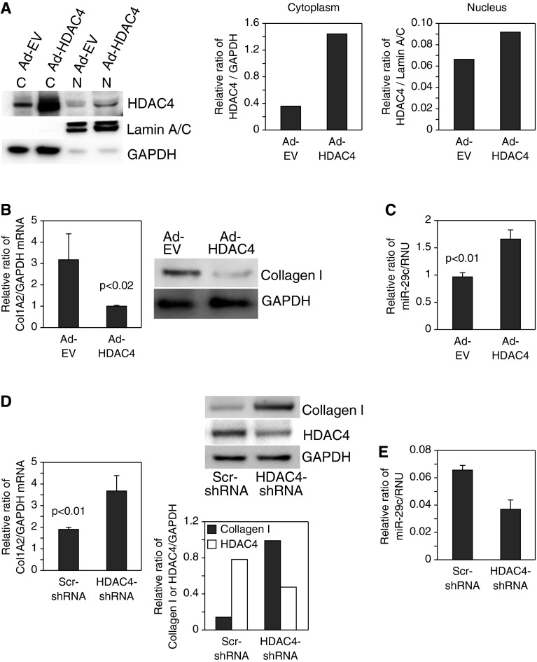
Figure 4.
HDAC4 regulates type I collagen and miR-29 expression. (A–C) HDAC4 was overexpressed in IPF fibroblasts using an adenoviral vector containing a wild-type HDAC4 construct (Ad-HDAC4). Controls consisted of cells expressing empty vector (Ad-EV). (A) Cells were seeded on polymerized type I collagen matrices for 4 hours. Cells were lysed and nuclear (N) and cytoplasmic (C) fractions were analyzed for HDAC4 protein levels by Western analysis (left panel). Nuclear and cytoplasmic HDAC4 levels were quantified by densitometric analysis (middle and right panels). (B) COL1A2 messenger RNA (mRNA) (left panel) and collagen I protein (right panel) expression were examined by qRT-PCR and Western blot analysis, respectively. GAPDH is shown as a loading control. (C) miR-29c levels were quantified by qRT-PCR. (D and E) HDAC4 was knocked down in control fibroblasts using a lentiviral vector containing HDAC4 short hairpin RNA (shRNA). Cells infected with lentiviral vector containing scrambled shRNA (Scr-shRNA) were used as a control. The cells were plated on polymerized type I collagen for 4 hours. (D) COL1A2 mRNA (left panel) and collagen I and HDAC4 protein levels (top right panel) were examined by qRT-PCR and Western blot analysis, respectively. GAPDH is shown as a loading control. Collagen I and HDAC4 protein levels were quantified by densitometric analysis (bottom right panel). (E) miR-29c levels were quantified by qRT-PCR.

