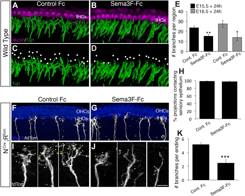
Figure 7. Exogenous Sema3f inhibits SGN outgrowth in vitro.
(A–D) Three-dimensional renderings of confocal z-stacks from cultured cochleae treated with either control IgG-Fc or Sema3F-Fc at 20 nM. The cultures were stained with anti-myosin VI to show the position of the HCs (magenta) and Tuj1 to mark SGNs (green). By comparison with an Fc-treated control (A, C) in which the SGNs show extensive branching, Sema3F-Fc-treatment significantly inhibited branching (B, D). The white dots in C and D mark individual branch points. (E) Quantification of SGN branch numbers normalized to the length of the cochlea within the photographed region. For this normalization, we measured the length of the cochlea along the IHC border. Addition of Sema3F-Fc led to significantly fewer SGN branches in explants initiated at E15.5 (black bars) or E16.5 (gray bars). **p ≤ 0.01; *p ≤ 0.05. n = 6 cochleae per treatment group. (F–K) To determine whether Sema3F-Fc reduced the number of SGNs in the cochlear epithelium or reduced the number of secondary branches per SGN, the Sema3F-Fc experiments were repeated using cochleae from Neurog1CreERT2; R26R-tdTomato mice. (F, G) Low-magnification micrographs from cochleae stained with anti-myosin VI (HCs, blue) and tdTomato to mark individual SGNs (white). (H) There were no differences in the percentages of labeled SGNs contacting the cochlear sensory epithelium in either group. (I, J) High-magnification images of representative individually labeled SGN terminals. Yellow dots mark small secondary branches extending from each SGN peripheral process. Note the reduced number of secondary branches in Sema3F-Fc-treated explants. (K) Quantification of the number of secondary branches per SGN ending showing the inhibitory effect of Sema3F-Fc. ***p ≤ 0.001. n = 6 cochleae per treatment group. Error bars, sem. Scale bar in J: 15 μm, A–D; 30 μm, F and G; 7 μm, I and J.

