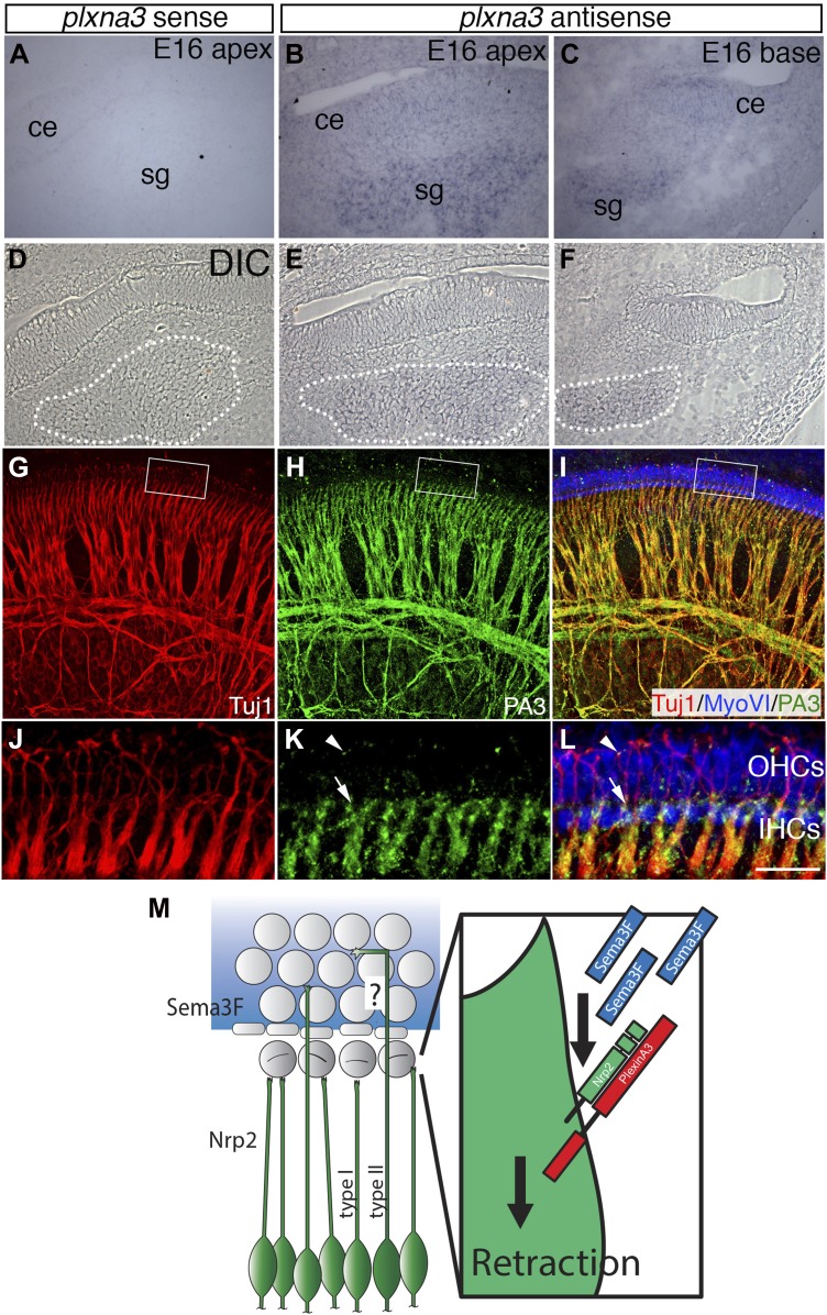
Figure 9. Differential expression of Plexin-A3 in SGNs and a proposed model.
(A–F) In situ hybridization for Plxna3 at E16.5. (A) A Plxna3 sense control probe does not show any reactivity. (B, C) Plxna3 transcripts are present in the SG, and possibly faintly in cells throughout the cochlear epithelium (ce). (D–F) Differential interference contrast (DIC) images for A–C were used to identify SGNs by morphology. The white dotted lines encircle the SGNs in each panel. (G–I) Plexin-A3 antibody staining in an E16.5 whole-mount cochlea. (G–I) Expression of Plexin-A3 (green) in SGNs appears to overlap with a neuronal marker (Tuj1 in red) except in the HC region (MyoVI staining in blue). (J–L) High-magnification views from the boxed regions in G, H, and I. Plexin-A3 is strongly expressed in SGN peripheral processes located adjacent to the IHC region (arrows) but appears markedly reduced in fibers that cross into the OHC region. Limited Plexin-A3 is detectable on sparse numbers of SGN processes in the OHC region (arrowhead). Scale bar in L: 100 μm, A–I, 25 μm, J–L. (M) Proposed model: Sema3F (blue shading and blue rectangles) is expressed by cells in the OHC region of the cochlea with a sharp boundary along the PCs. Binding of Sema3F to Nrp2 (green SGNs and green rectangle) induces retraction events that inhibit the growth of the type I SGN peripheral processes into the OHC domain. PlexinA3 co-receptors, which likely serve a role in this process, are shown in red. ‘?’ illustrates an outstanding question of how type II SGNs, which express Nrp2 and are confronted by Sema3F, are able to extend into the OHC domain.


