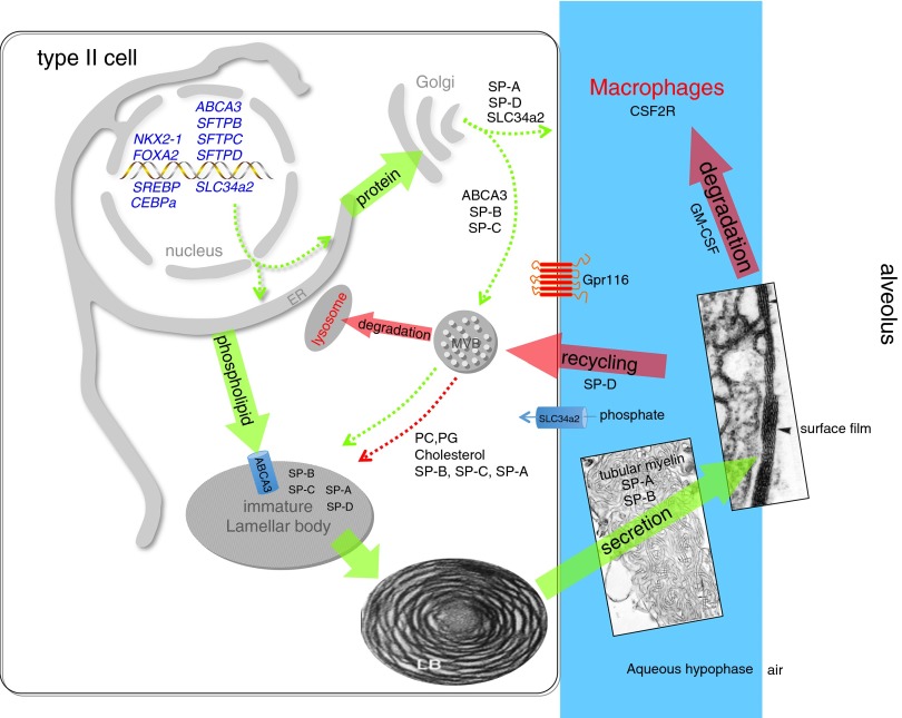
Figure 4.
Surfactant metabolism. NKX2–1 (TTF-1) is a transcription factor critical for differentiation of type II epithelial cells and regulation of expression of Abca3, Slc34a2 (a phosphate transporter), and the surfactant proteins. Newly translated surfactant proteins (SPs) (proSP-B and proSP-C) and lamellar body proteins (ABCA3) traffic from the endoplasmic reticulum (ER) to the Golgi and subsequently to the multivesicular body (MVB); in contrast, SP-A and SP-D are constitutively secreted by the type II epithelial cells (dotted green arrows). Fusion of the MVB with the lamellar body (LB) is accompanied by proteolytic processing of SP-B and SP-C proproteins to their mature peptides. Newly synthesized surfactant phospholipids (DPPC, PG) are likely transported directly from the ER to the LB by lipid transfer proteins. The contents of the LB are secreted into the alveolar space, where they interact with SP-A to form tubular myelin and, ultimately, a phospholipid-rich film (surfactant) at the air–liquid interface. Cyclical expansion and compression of the bioactive film result in incorporation (green arrow) and loss (red arrows) of lipids/proteins from the multilayered surface film. Alveolar surfactant lipids and proteins are cleared through a granulocyte/macrophage colony-stimulating factor–dependent pathway that regulates alveolar macrophage differentiation and function. Surfactant remnants are also taken up by the type II epithelial cell and recycled to the LB via the MVB for resecretion, and a portion is degraded in lysosomes. SP-D plays an important role in regulating alveolar surfactant pool size, likely by enhancing its reuptake by type II epithelial cells; Gpr116 also regulates alveolar surfactant pool size by an unknown mechanism. The MVB integrates surfactant synthesis, secretion, recycling, and degradation pathways in the type II cell. Green arrows indicate biosynthetic pathways; red arrows indicate degradation and recycling pathways. Dotted arrows represent trafficking routes of proteins and lipids.

