
Figure 2. FFP imaging of an acrylic phantom filled with six populations of SPIO-labeled cells.
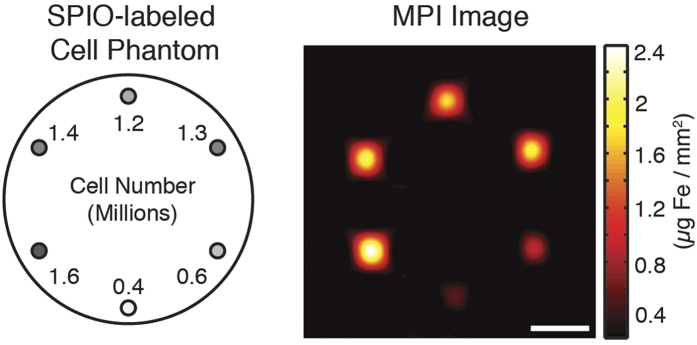
The MPI signal from cell populations (ranging from 4 × 10⁵ to 1.6 × 10⁶ in number) corresponds linearly to iron oxide tracer concentration, enabling quantitative imaging. Imaging parameters: 3.5 min acquisition on a 7 T/m 3D MPI scanner, 5 cm × 4.5 cm × 3 cm FOV. Scale bar: 1 cm.