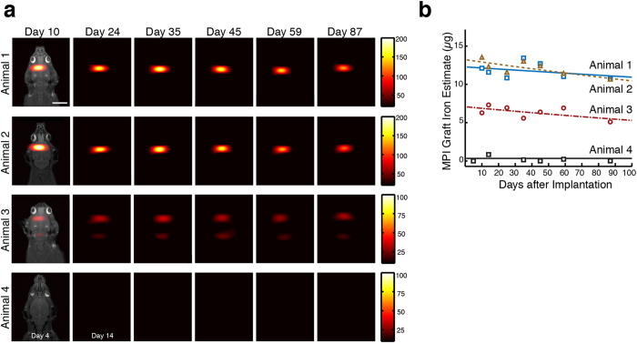
Figure 3. MPI quantitatively tracks NPC neural implants in rats over 87 days.
(a) Longitudinal MPI imaging of 5 × 10⁵ SPIO-labeled human NPCs implanted in the forebrain cortex (Animals 1-2), near lateral ventricle (Animal 3), and equivalent SPIO-only tracer in the forebrain cortex as control (Animal 4). MPI imaging quantifies graft clearance and movement over time, with rapid total clearance of SPIO-only injection (MPI: 30 sec acquisitions, 9.3 cm × 6 cm FOV, color intensity in ng/mm2, MRI reference: 10 min acquisitions, 7.5 cm × 4 cm, 384 × 384 matrix). All images are equally scaled; scale bar: 1 cm. (b) Total iron MPI estimates for in vivo cell grafts are plotted as a function of time with exponential fit. In vivo iron in Animal 1 and 2 do not show significant decrease over time, while iron signal was significantly decreased in Animal 3 starting Day 10. Animal 4 showed no long-term persistent MPI signal.

