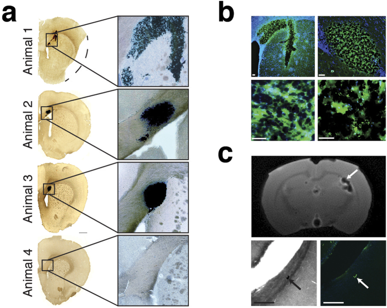
Figure 4. Histological and MRI validation of iron location and quantification.
(a) Histological analysis of NPC grafts. Postmortem Prussian blue (PB) staining confirms presence of iron-labeled cells at administration site but not for SPIO-only control (Animal 4). (b) Representative immunohistochemical slices (top right panels) are shown for NPC marker nestin (top left, Animal 1), neural cell adhesion molecule (top right, Animal 3), and human-specific cytoplasmic marker SC121 (bottom left, Animal 2), which indicate iron label within NPC grafts. CD68 staining (bottom right, Animal 3) also indicates immune cells at administration site, suggesting immune-based graft clearance. Scale bars: 100 μm. (c) Postmortem axial MRI indicates iron in lateral ventricle in Animal 3 (arrow). PB and CD68 staining of lateral ventricle in Animal 3 shows SPIO uptake by immune cells. MRI: 20 min acquisition, 2.56 cm isotropic FOV, 2563 pixel matrix. Scale bars: 1 mm.

