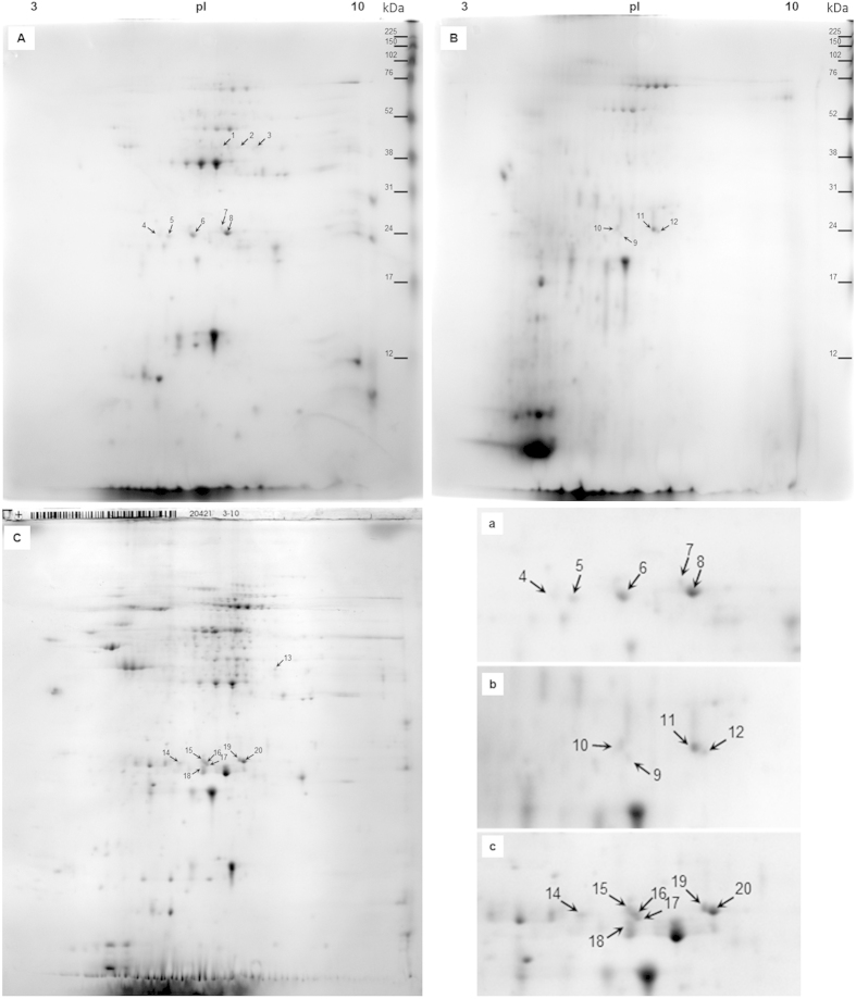
Figure 2. 2D-E 14% pI 3-10 analysis of protein fractions from Varroa destructor with localized virus structural proteins.
Legend: (A) p-ABA-unbound fraction, (B) p-ABA-purified fraction, (C) supernatant-before-purification (e.g., total soluble proteome). Lowercase marked figures (a–c) show the details of ~23–24 kDa viral spots in the corresponding fractions. For details on the viral spots identified using MALDI TOF/TOF, see Table 2; and for details on MS/MS identification, see Table Supplement 2.

