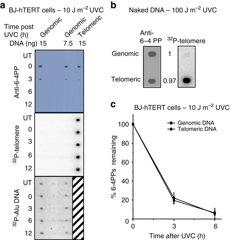
Figure 3. Quantification of 6–4PP formation and removal in telomeres from UVC exposed BJ-hTERT cells.
(a) Cells were untreated or exposed to 10 J m−2 UVC followed by collecting at various repair times (0–12 h). Telomeres were isolated from purified genomic DNA (100 μg each time point) and combined from two separate experiments to obtain 15 ng (lane 3) for loading. Genomic DNA was loaded at 15 ng (lane 1) and 7.5 ng (lane 2). The blot was sequentially probed with a 6–4PP antibody, a radiolabelled telomere probe, and a radiolabelled Alu repeat probe. (b) Purified genomic DNA (100 μg) was exposed to 100 J m−2 UVC in vitro. Telomeres were isolated and blotted (7 ng) with equal amounts of genomic DNA and probed with the 6–4PP antibody and radiolabeled telomere probe. The 6–4PP signal intensity was quantitated and normalized to genomic DNA for comparison; from three independent experiments. (c) The 6–4PP signal intensity was quantitated for experiments in a, normalized to 0 h, and plotted against recovery time. Values and error bars are the mean and s.e.m. from four independent experiments for genomic DNA and two experiments for telomeric DNA. The difference between the curves was not statistically significant (P=0.95) by two-way ANOVA.

