
FIGURE 5.
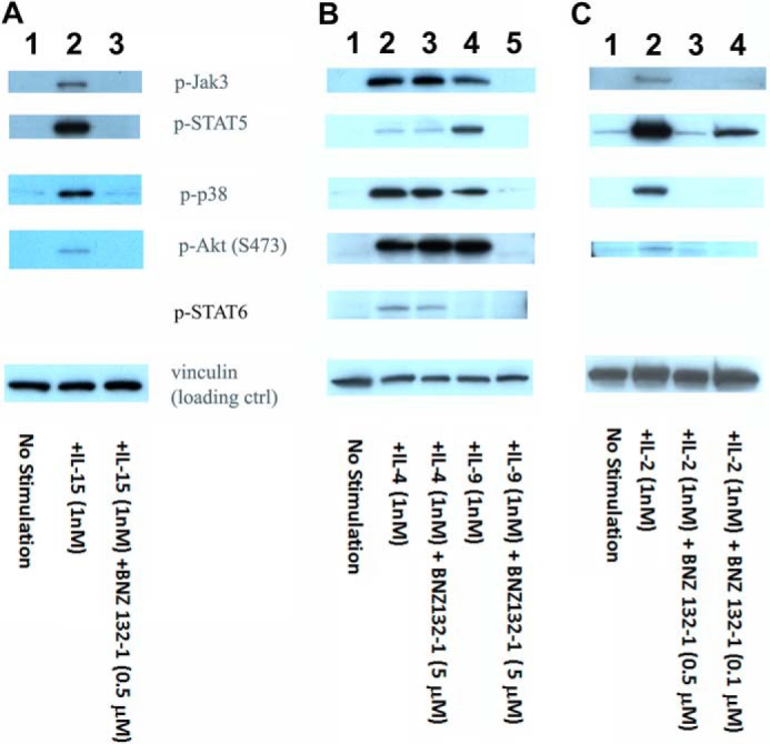
Comprehensive inhibition by BNZ132-1 of signaling events caused by target γc-cytokines. A, comprehensive inhibition of IL-15 signaling in PT-18β by BNZ132-1. Cells had been withdrawn from IL-3 for 12 h and then stimulated with 1 nm human IL-15 with or without 0.5 μm BNZ132-1 for 15 min before extracting cellular proteins. Phosphorylation of targets was detected using commercial antibodies (Cell Signaling), followed by ECL. Lane 1, no stimulation; lane 2, IL-15(1 nm); and lane 3, IL-15 (1 nm) + BNZ132-1 (0.5 μm). B, signal transduction of IL-4 in PT-18 was not inhibited, whereas the IL-9 signal was inhibited by BNZ132-1. Cells were fasted from IL-3 for 12 h then stimulated with 1 nm mouse IL-4 or mouse IL-9 in the presence or absence of an excess dose of BNZ132-1 (5 μm) for 15 min before extracting cellular proteins. Phosphorylation of the indicated proteins was detected using phosphospecific antibodies (Cell Signaling), followed by ECL. IL-4 is the only γc-cytokine that induces the phosphorylation of STAT6. As shown before, only IL-4, but not IL-9, induced the tyrosine phosphorylation of STAT6. Conversely, IL-4 showed only marginal phosphorylation of STAT5, whereas IL-9 induced a strong STAT5 phosphorylation. Notably, only IL-9 action was inhibited by BNZ132-1. Lane 1, no stimulation; lane 2, IL-4; lane 3, IL-4 + BNZ132-1; lane 4, IL-9; and lane 5, IL-9 + BNZ132-1. C, IL-2 signal in human PBMC was blocked by BNZ132-1. Human ex vivo T cells were prepared as described in the text. After 24 h of IL-2 depletion, cells were stimulated by 1 nm IL-2 with or without BNZ132-1 (0.5 or 0.1 μm) for 20 min before lysis. Tyrosine phosphorylation of targets was detected by antibodies (Cell Signaling), followed by ECL. Lane 1, no stimulation; lane 2, IL-2 stimulation; lane 3, IL-2 + BNZ132-1 (0.5 μm); and lane 4, IL-2 + BNZ132-1 (0.1 μm). In all blots, anti-vinculin Ab (Sigma) was used to validate equal protein loadings.