FIGURE 2.
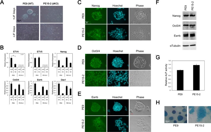
Etv4/5 dKO ES cells express self-renewal marker genes. A, morphological signatures of PE9 (WT) and PE15-2 (Etv4/5 dKO) ES cells. PE9 and PE15-2 ES cells were cultured in the presence or absence of LIF for 3 days. Morphological signatures were observed. B, expression of self-renewal marker genes in PE9 and PE15-2 ES cells. Expression of the indicated self-renewal marker genes in the conditions of A in PE9 and PE15-2 ES cells was examined by quantitative RT-PCR. Expression of Etv4 and Etv5 mRNA was only shown in PE9 ES cells. All samples were analyzed in triplicate, and the data were normalized to Gapdh expression. C–E, immunofluorescent stain in PE9 and PE15-2 ES cells. Protein expression of the self-renewal marker genes, including NANOG (C), OCT3/4 (D), and ESRRB (E), in PE9 and PE15-2 ES cells was examined by immunofluorescent stain. Nuclei were stained with the Hoechst stain. F, Western blot analysis in PE9 and PE15-2 ES cells. Protein expression of the self-renewal marker genes (NANOG, OCT3/4, and ESRRB) was examined by Western blot analysis. α-Tubulin was used as a loading control. G, determination of alkaline phosphatase activity. Relative activity of alkaline phosphatase was determined in PE9 and PE15-2 ES cells. Shown are the means (bars) and S.D. (error bars) of triplicate plates. H, alkaline phosphatase stain in PE9 and PE15-2 ES cells. Alkaline phosphatase of ES cells was detected by the VECTOR Blue alkaline phosphatase substrate kit. Morphological signatures were observed. All results are representative of several independent experiments.

