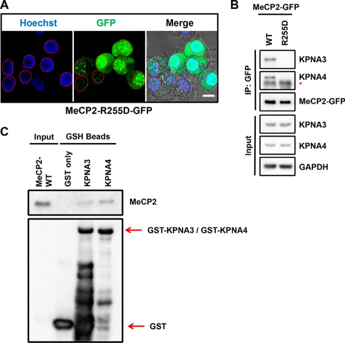
FIGURE 4.
The MeCP2-R255D mutant disrupts nuclear localization and the interaction with KPNA3 and KPNA4. A, N2a cells were transiently transfected with a human MeCP2-R255D construct tagged on the C terminus with GFP and live-imaged using endogenous GFP excitation with Hoechst 33342 as a nuclear counterstain. Individual channels are indicated, and a merged image overlaid on the DIC channel is shown to indicate nuclear and cytoplasmic boundaries. A dashed red line indicates the nuclear outline of each transfected cell. Scale bars = 10 μm; n = 3. B, N2a cells were transiently transfected with either MeCP2 WT or MeCP2-R255D. Two days later, cells were lysed, and MeCP2 was isolated by precipitation with anti-GFP antibodies. Bound proteins were eluted and identified by Western blotting with the indicated antibodies, along with an aliquot of the input protein. Asterisk, the location of the immunoglobulin heavy chain band below the KPNA4 band. n = 3. C, GSH beads loaded with bacterially expressed GST-KPNA3, GST-KPNA4, or GST only were incubated with bacterially expressed MeCP2-WT in vitro. Bound proteins were eluted and identified by Western blotting with the indicated antibodies, along with an aliquot of input MeCP2 protein. n = 3.

