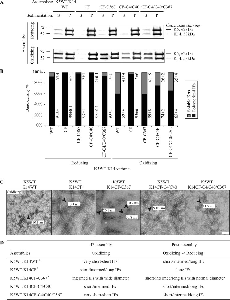
FIGURE 4.
Comparison of K14WT-, K14CF-, K14CF-C367-, K14CF-C4/C40-, and K14CF-C4/C40/C367-containing filaments assembled in vitro under either reducing or oxidizing buffer conditions. A, high-speed sedimentation assay (150,000 × g, 30 min) followed by SDS-PAGE analysis of supernatant (S) and pellet (P) fractions. B, quantitation of polymerization efficiency using densitometry-based analysis of fractions shown in A. Data are presented as average ± S.D. from three independent experiments. C, negative staining and transmission electron microscopy were performed to compare the ultrastructural features of the keratin pairing shown on the top of each micrograph. Diameters of representative individual filaments (denoted by arrowheads) assembled in vitro under oxidizing buffer conditions are labeled in each micrograph. Bar, 100 nm. D, summary of in vitro assembly data for various keratin combinations under different buffer conditions. For the K5WT/K14WT, K5WT/K14CF, and K5WT/K14CF-C367 pairings, similar findings were originally reported in Ref. 17. We repeated these analyses in the current study in order to carry out a comparison with the K5WT/K14CF-C4/C40 and K5WT/K14CF-C4/C40/C367. Very short IFs, <150 nm; short IFs, 200–500 nm; Intermed. IFs, 500–1000 nm; long IFs, >1 μm.

