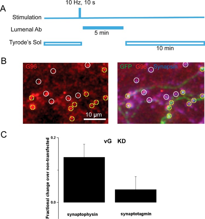
FIGURE 2.
vGlut deficiency slows the synaptic activity-driven recycling of endogenous synaptophysin but synaptotagmin. A, illustration of the experimental protocol (see “Experimental Procedures” for details). Cells were stimulated for 10 s at 10 Hz and followed 10 s after the end of the stimulus by perfusion with a luminal antibody either against synaptophysin (G96 serum) or against synaptotagmin 1 (Oyster-550-labeled anti-Syt1). After a 5-min incubation, the unbound antibody was washed out in Tyrode's solution (Tyrode's Sol) for 10 min, fixed, permeabilized, and stained with secondary and other antibodies. B, a representative image of G96 serum-labeled boutons (left) and its overlay image with GFP in green and synapsin I in blue (right). GFP-positive boutons (yellow circles) and GFP-negative boutons (white circles) were selected based on positive synapsin I staining when the luminal antibody channel was blinded. C, for each randomly selected imaging field containing both GFP-positive and GFP-negative boutons, the fluorescence intensity of the lumenal antibody at the GFP-positive bouton was normalized to the averaged fluorescence intensity of control boutons in the same field. VGlut1 deficiency (vG KD) resulted in 13.7 ± 4.4% increase in synaptophysin (Syphy) staining (n = 12 cells, 224 boutons) when compared with the untransfected control (n = 12 cells, 215 boutons, p = 0.026, paired t test). In contrast, vG KD did not change the synaptotagmin 1 (Syt1) staining (4.1 ± 4.3% increase, n = 11 cells, 125 boutons) relative to its control (n = 11 cells, 163 boutons, p = 0.42, paired t test). Error bars indicate S.E.

