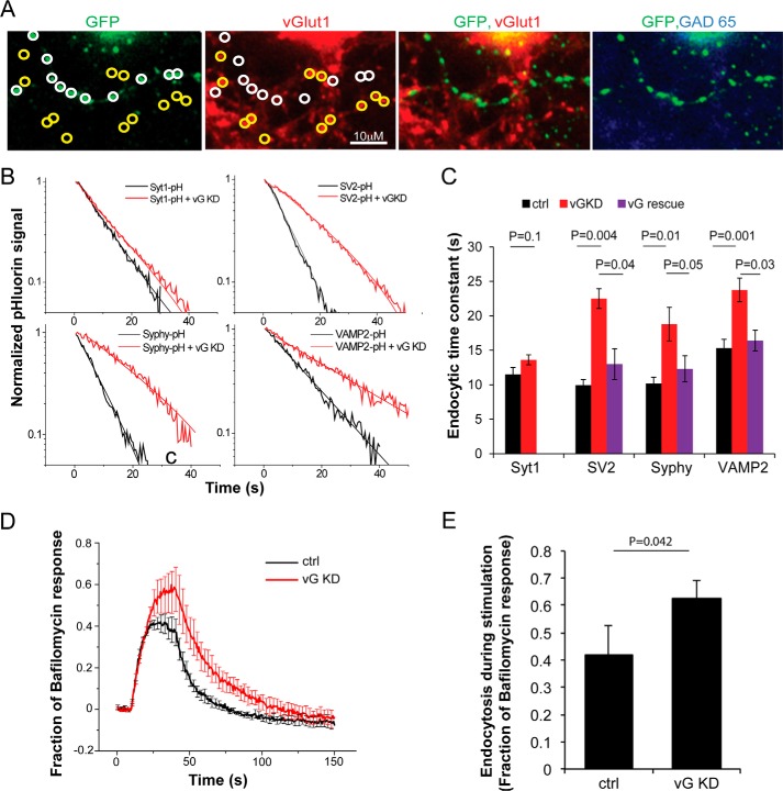
FIGURE 3.
vGlut modulates endocytic retrieval of 3 different SV cargos at nerve terminals. A, representative presynaptic boutons from a neuron co-expressing a pHluorin-tagged SV protein (stained with anti-GFP) and vGlut1 shRNA. GFP-positive boutons (white circles) show much lower vGlut1 immunofluorescence when compared with neighboring non-transfected boutons (yellow circles). Only neurons that were negative for anti-GAD65 immunoreactivity (right panel) were used in this study (positive GAD-65 puncta were visible on the same coverslip, although not in this field of view). B, decay of SV-pHluorin fluorescence after a 10-s 10 Hz stimulation showing endocytosis kinetics (thick traces) in semi-log format with single-exponential fits (thin lines) measured in vGlut1 knockdown (vG KD, red) and control (black) synapses. C, summary of endocytic time constants for different SV proteins in control (black), vG KD neurons (red), and those expressing shRNA-resistant vG on KD background (vG rescue, purple). The slowed kinetics observed for SV2, Syphy, and VAMP2 was rescued (SV2 rescue, 13.0 ± 2.2, n = 6 when compared with KD: 22.5 ± 1.4 s, n = 9, p = 0.04; Syphy rescue, 12.3 ± 1.9 s, n = 8 when compared with KD: 18.8 ± 2.4 s, n = 8, p = 0.05; VAMP2 rescue, 16.4 ± 1.5, n = 6 when compared with KD: 23.7 ± 1.7 s, n = 11, p = 0.03, Student's t test) by re-expression of vGlut1. D, averaged Syphy-pHluorin traces for a longer stimulus train (10 Hz, 30 s) are shown for WT (control (ctrl), n = 6) and neurons co-expressing vGLUT1 shRNA (vG KD, n = 7). Error bars indicate S.E. E, summary of total fraction of endocytosis during stimulation determined by subtracting the peak response to 10 Hz, 30 s stimulation without bafilomycin (as in D) from the response to the same stimulus in the presence of bafilomycin. Ctrl: 0.42 ± 0.11 when compared with KD: 0.63 ± 0.06, p = 0.04, Student's t test.

