
FIGURE 6.
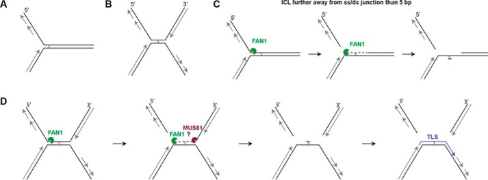
The putative mechanism of FAN1-dependent ICL unhooking. A, schematic of a single replication fork arrested at an ICL. B, the X-shaped structure arising through the convergence of two replication forks at an ICL. C, a single fork arrested at an ICL that is more than 5 bp from the ss/ds boundary would be incised by FAN1 5′ from the ICL, which would release the lagging strand. The enzyme would then degrade the nicked strand in a 5′-to-3′ direction to generate a substrate for translesion polymerases and subsequent repair by nucleotide excision repair. D, an X-shaped structure that contains a duplex longer than 10 bp where the lagging and leading strand flaps could be released by the action of FAN1 and, e.g., MUS81.