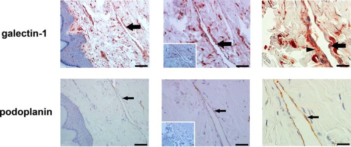
FIGURE 1.

Human lymphatic endothelial cells express and secrete galectin-1. Sections of skin from lymphedema patients were stained with polyclonal antibody against galectin-1 (top) or monoclonal antibody to the human LEC marker podoplanin (bottom). Bound antibody was detected with the corresponding secondary antibody and visualized using a 3-amino-9-ethylcarbazole chromogenic substrate system. Sections were counterstained with hematoxylin. Insets (middle column) show control antibody staining. Dilated lymphatic vessels are lined by LECs expressing galectin-1 (arrow, top) and podoplanin (arrow, bottom). Data are representative of six independent tissue samples. Note that the distribution of galectin-1 on LECs appears more dispersed than that of podoplanin, suggesting the localization of secreted galectin-1 in extracellular matrix (arrowhead, top right panel). Magnification is as follows: ×20 (left), ×40 (middle), and ×100 (right). Scale bar, 100 μm (left), 50 μm (middle), and 20 μm (right).
