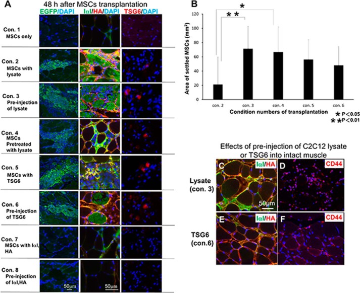
FIGURE 2.
Morphological analysis 48 h after transplantation of MCSs into noninjured skeletal muscle under various conditions. Eight different conditions for the transplantation are shown in Table 1. A, expression of EGFP in green (left column) indicates the MSCs transplanted. Middle column shows expression of IαI (green) and HA (red) in each condition (Con.). Right column shows TSG6 (red) immunoreactivity in each condition. B, area where MSCs settled in each condition were calculated and compared with each other. Under conditions 3 and 6 (see A and Table 1) where MSCs were successfully transplanted into the intact muscle tissues, pre-injection of C2C12 lysate (C and D) and TSG6 (E and F) increased the expression of IαI (green) and HA (red) (C and E) and CD44 (red) (D and F) after 6 h. MSCs transplanted were found to settle in those areas where IαI and HA surrounded muscle fibers and co-localized with each other. Scale bar in A, left bottom panel, corresponds to all panels in left column. Scale bar in A, middle bottom panel, corresponds to all panels in middle and right columns. Scale bar in C corresponds to C–F (n = 3).

