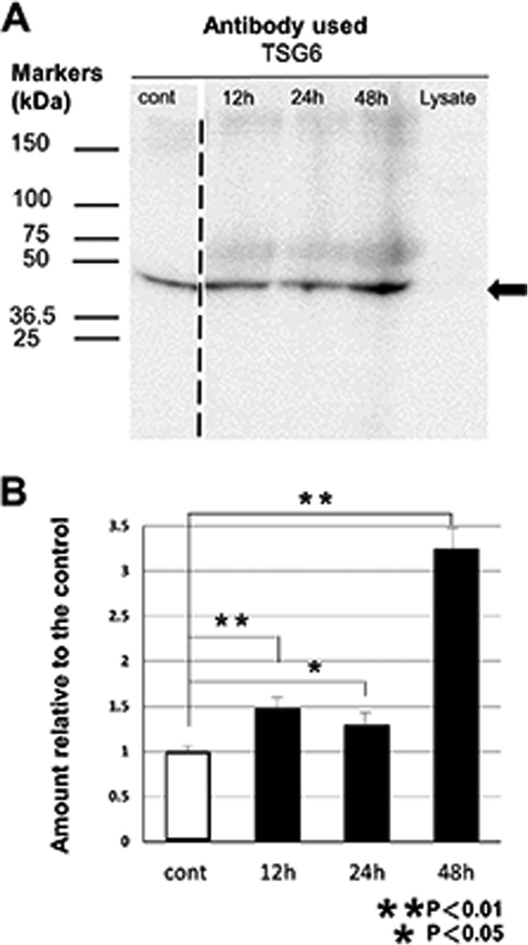
FIGURE 4.
Protein TSG6 released into medium from MSCs by stimulation with C2C12 lysates. Conditioned medium samples for 12, 24, and 48 h were obtained from MSC cultures incubated for 12, 24, and 48 h, respectively, in medium containing 0.5 mg/ml (protein concentration) lysate of C2C12. The medium sample for the control (Cont) was obtained from the culture incubated in normal medium for 48 h. Western blotting analysis for equal amounts of proteins (20 μg) obtained from these conditioned media revealed the presence of TSG6 protein in MSC-conditioned medium (A). Lysate of C2C12 did not contain detectable TSG6, but after the addition of C2C12 lysate, protein TSG6 in medium of MSC culture increased. Arrow indicates the migrating position of recombinant TSG-6 from R&D Systems. Faint and broad bands with the lower migrating positions than the recombinant TSG-6 may correspond to TSG-6-HCs and TSG-6-IαI and their degraded products (44, 49, 50). The broken line between the lanes for the control and for the 12-h cultured medium indicates that there were two lanes in the original figure that contained different amounts (2- and 3-fold) of proteins derived from the control medium cultured for 48 h, and now they were deleted to avoid redundancy. Densities of the probed bands were analyzed using ImagePro software, and integrated densities were normalized to the control medium (mean ± S.D., n = 3) (B).

