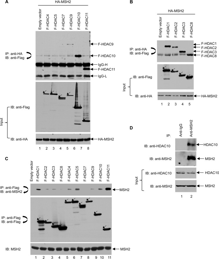
FIGURE 1.
Multiple HDACs interact with MSH2. A, several Class II HDACs interact with MSH2 in 293T cells. 293T cells were transiently transfected with plasmids expressing HA-MSH2 and FLAG (F)-tagged Class II HDACs as indicated. HA-MSH2 immunoprecipitation (IP) with anti-HA-agarose beads was performed followed by immunoblotting (IB) with an anti-FLAG antibody (upper panel). Total cell lysates were subjected to immunoblot analyses with anti-FLAG and anti-HA antibodies (middle and lower panels). B, all Class I HDACs interact with MSH2 in 293T cells. 293T cells were transiently transfected with plasmids expressing HA-MSH2 and FLAG-tagged Class I HDACs as indicated. HA-MSH2 immunoprecipitation with anti-HA-agarose beads was performed followed by immunoblotting with an anti-FLAG antibody (upper panel). Total cell lysates were subjected to immunoblot analyses with anti-FLAG and anti-HA antibodies (middle and lower panels). C, multiple HDACs interact with endogenous MSH2 in 293T cells. 293T cells were transiently transfected with plasmids expressing the indicated FLAG-tagged Class I and II HDACs. Immunoprecipitation of HDACs with anti-FLAG M2-agarose beads was performed followed by immunoblot analyses with anti-MSH2 antibody to detect endogenous MSH2 (upper panel). The blot was then stripped and reprobed with anti-FLAG antibody (middle panel). Levels of MSH2 were confirmed by Western blot analysis of the cell lysates using an anti-MSH2 antibody (lower panel). D, interaction between endogenous HDAC10 and MSH2 in HeLa cells. HeLa cell lysates were first immunoprecipitated with anti-IgG or anti-MSH2 antibody and then subjected to immunoblot analysis using an anti-HDAC10 antibody. The blot was then stripped and reprobed with an anti-MSH2 antibody.

