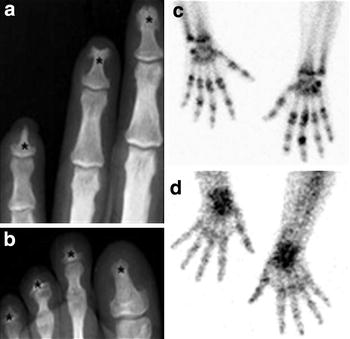
Fig. 4.

Newly diagnosed lepromatous patients exhibits bone and cartilage lesions. a, b Radiography of left hand and foot. Asterisks indicate the distal phalanx erosion, typical hallmark of leprosy. c Third-phase bone scintigraphy image shows increased bone uptake of 99mTc-MDP in both hands. Hyper-fixation occurred in bones and joints of phalanges, metacarpus and wrists. Important joint alterations were evidenced in all patients. d The early phase scintigraphy shows localised radiotracer activity on the wrists
