Figure 1.

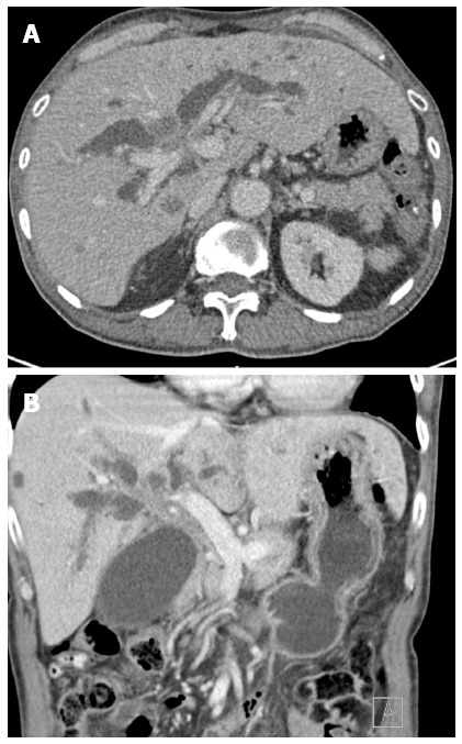
Computed tomography examination. A: Axial; B: Coronal computed tomography images showing an ill-defined mass in the hilar region causing bilateral intrahepatic duct dilation.

Computed tomography examination. A: Axial; B: Coronal computed tomography images showing an ill-defined mass in the hilar region causing bilateral intrahepatic duct dilation.