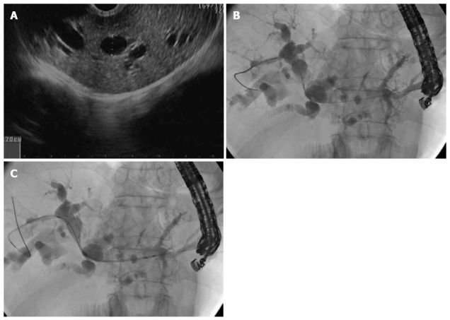
Figure 2.

Guidewire insertion. A: Echoview demonstrating dilated of left system B: Cholangiogram showing the tapered-tip sphincterotome; C: The guidewire was passed to the right system from the gastric site and an uncovered self-expanding metal stent was deployed.
