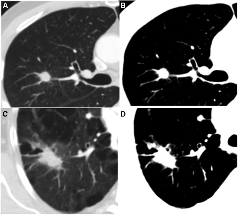
Figure 2.
Axial computed tomography images from two cases with non–small cell lung cancer. (A) A 1.1-cm lesion in a region of 1.6% low-attenuation areas less than −950 Hounsfield units (%LAA−950). Each case was opened into a “blinded” window setting (B) before measurement of the tumor. (C) A 3.9-cm right upper lobe lesion. The tumor %LAA−950 in this region was 24.6%. The “blinded” window setting (D) effectively obscures even large amounts of emphysema, allowing measurement of tumor diameter in an objective manner.

