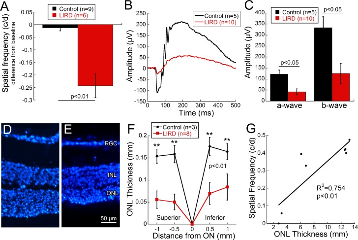
Figure 2.
Behavioral, functional, and histologic analyses confirmed light-induced retinal damage in pigmented animals. Visual acuity decreased significantly from baseline in LIRD animals 6 days post light exposure ([A] Student's t-test = 3.79, P < 0.01). Averaged waveform (B) in response to bright-flash (4.1 cd s/m2) stimulus from CTRL (black) and LIRD (red) 129/SvJ mice at 7 days post LIRD. Significant deficits in the absolute value of the a- and b-wave response occurred between CTRL and LIRD animals ([C] a-wave: Mann-Whitney rank sum test = 4.0, P = 0.01, b-wave: Mann-Whitney rank sum test = 5.0, P = 0.02). Measurements of DAPI-stained retinal cross sections confirmed thinning of the outer nuclear layer (ONL) thickness between CTRL (D) and LIRD (E) animals as quantified in (F). The ONL was significantly thinner in the LIRD mice compared to controls (2-way repeated ANOVA F(3, 43) = 4.68, P = 0.009). Visual acuity and ONL thickness are strongly correlated ([G] correlation R2 = 0.754, P < 0.01), with reduction in ONL thickness corresponding to decreased visual acuity. Data shown are mean ± SEM. Asterisks represent significant post hoc comparisons, **P < 0.01. RGC, retinal ganglion cell layer; INL, inner nuclear layer.

