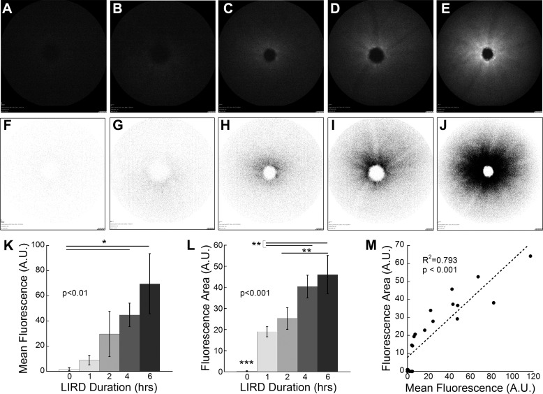
Figure 4.
Representative fundus images of 129/SvJ mice exposed to increasing durations of light toxicity after intravenous H-800CW injections. Animals were subjected to 60,000 lux light for durations of 0 (CTRL; n = 7; [A, F]), 1 (n = 3; [B, G]), 2 (n = 4; [C, H]), 4 (n = 4; [D, I]), and 6 (n = 3; [E, J])) hours. Mean fluorescence was measured (K) from the images obtained with SLO (A–E). Animals subjected to light damage for 4 and 6 hours yielded mean fluorescence values that were significantly greater than in CTRL animals ([K] Kruskal-Wallis 1-way ANOVA on ranks, H = 16.17, P = 0.003). Area of hyperfluorescence was measured from color inverted SLO images (F–J). Hyperfluorescence area was significantly greater in all mice exposed to toxic light compared to CTRL (0 hours; [L] 1-way ANOVA F(4, 20) = 21.68, P < 0.001). Additionally, significant differences were found between mice exposed to increasing toxic light, illustrating the sensitivity of H-800CW to detect ROS levels with increasing retinal damage. Quantification by area or intensity was highly correlated with area of hyperfluorescence showing greater sensitivity (M). Post hoc comparisons *P < 0.05, **P < 0.01, ***P < 0.001.

