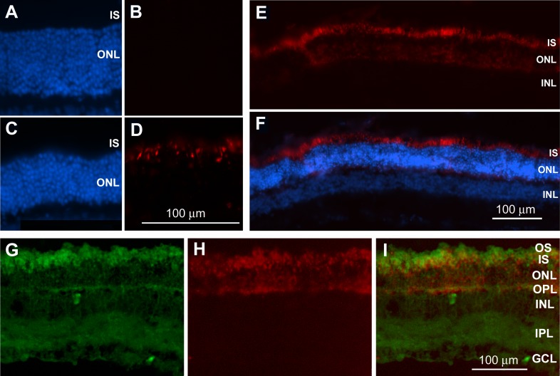
Figure 7.
Representative micrographs from retinal sections of Long Evans rats that were intravitreally injected with H-800CW (red) in vivo. DAPI staining (blue) in CTRL (A) and LIRD (C) retinas enabled visualization of retinal layers. Oxidized H-800CW fluorescence was absent from CTRL retinas (B) but strongly labeled the inner segments (IS) and outer nuclear layer (ONL) of LIRD retinas (D). A lower-magnification image of the retina showing consistent ROS labeling localized within the IS and ONL (E, F). Dichlorofluorescein labeling ([G] green) of retinas from H-800CW-injected eyes ([H] red) shows colabeling in the photoreceptor inner segments, outer nuclear layer, and outer plexiform layer (I), confirming that oxidized H-800CW labels ROS. OS, outer segments; IS, inner segments; INL, inner nuclear layer; IPL, inner plexiform layer; GCL, ganglion cell layer.

