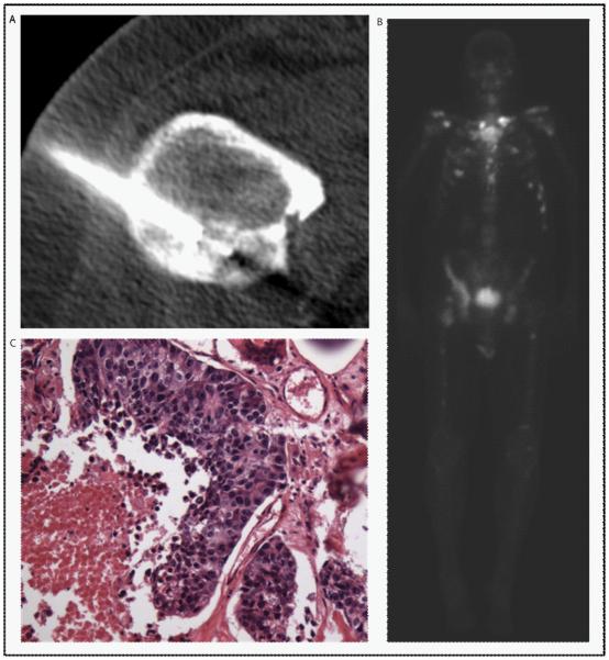
Figure 1.
CT (A), bone scan (B), and pathology (C) images from a CT-guided biopsy of a sclerotic, right lesser trochanter lesion, measuring 4.1 × 3.2 × 3.4 cm obtained at progression on abiraterone. The bone scan showed mild radiotracer uptake at the site of the lesion targeted for biopsy. The pathology was positive for tumor by light microscopy.

