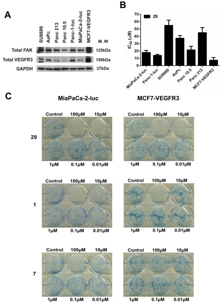
Fig. 5.
In vitro cytotoxicity profile of analog 29 in pancreatic cancer cell lines. (A) Analysis of FAK and VEGFR3 protein expression in six pancreatic cancer cell lines and MCF7-VEGFR3 cell line. GAPDH was used as a loading control. (B) IC50 values of 29 in the indicated cell lines. Error bars represent ±SD. (C) MiaPaCa-2-luc or MCF7-VEGFR3 cells were treated with 29 at the indicated concentrations. After 14 days, colonies were stained with 0.25% of 1, 9-dimethyl-methylene blue in 50% methanol for 1 h. Representative images from three independent experiments have been shown. (For interpretation of the references to colour in this figure legend, the reader is referred to the web version of this article.)

