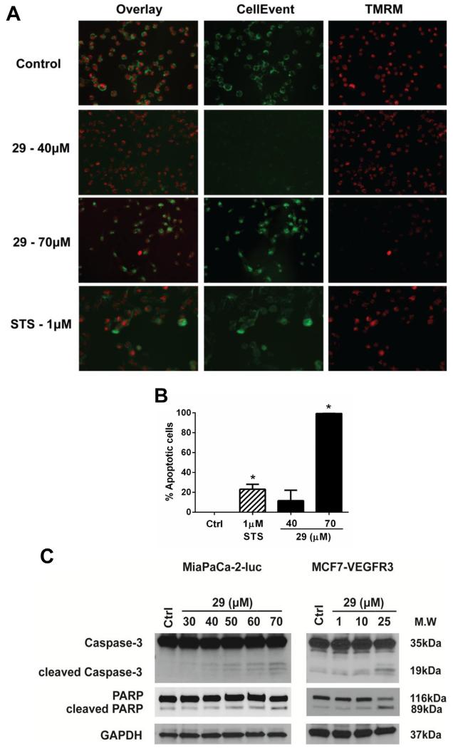
Fig. 7.
Analog 29 induced cell death via activation of the apoptotic pathway. (A) MiaPaCa-2-luc cells were treated with analog 29 at the indicated concentrations. After 24 h cells were stained with CellEvent™, a caspase-3, 7 dye and TMRM, a mitochondrial dye. Representative images are shown from three independent experiments. (B) Apoptotic cells were counted from multiple microscopic fields for each condition. Error bars represent ±SEM. *p < 0.05 (C) Addition of analog 29 to MiaPaCa-2-luc and MCF-VEGFR3 cells at the indicated concentrations for 24 h. Antibodies for caspase-3 and PARP were used to detect apoptosis. GAPDH serves as a loading control.

