Figure 4.
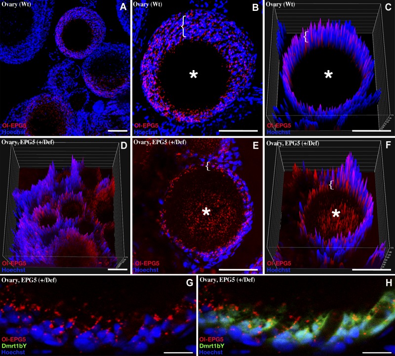
Ovarian localization of Ol-epg5 in wild-type and Ol-epg5-deficient fish. A–C) In the ovary, Medaka Ol-epg5 was expressed in the surrounding supporting cells (brackets) of the oocytes (asterisks). Ol-epg5 progressively accumulates in the supporting layer while the oocytes maturate (brackets in B and C). D–H) Ovarian localization of Ol-epg5 in a triple-transgenic fish (Ol-epg5-deficient/Ol-epg5:mCherry/Ol-dmrt1bY:GFP). In Ol-epg5-deficient ovaries, a clear accumulation of the Ol-epg5:mCherry fusion protein was apparent in the cell layer surrounding the oocytes (brackets) and in the oocytes (asterisks). C, D, F) Intensity surface projections show accumulation of epg5:mCherry fusion protein in the cytoplasm of oocytes (asterisk) of (D, F) Ol-epg5-deficient ovaries compared with wild type (C). G, H) In the surrounding layer of the oocytes, Ol-epg5 precisely colocalized with Ol-dmrt1bY, a marker of the granulosa cells (37, 38). Scale bars, 10 (A–H) and 40 µm (I, J).

