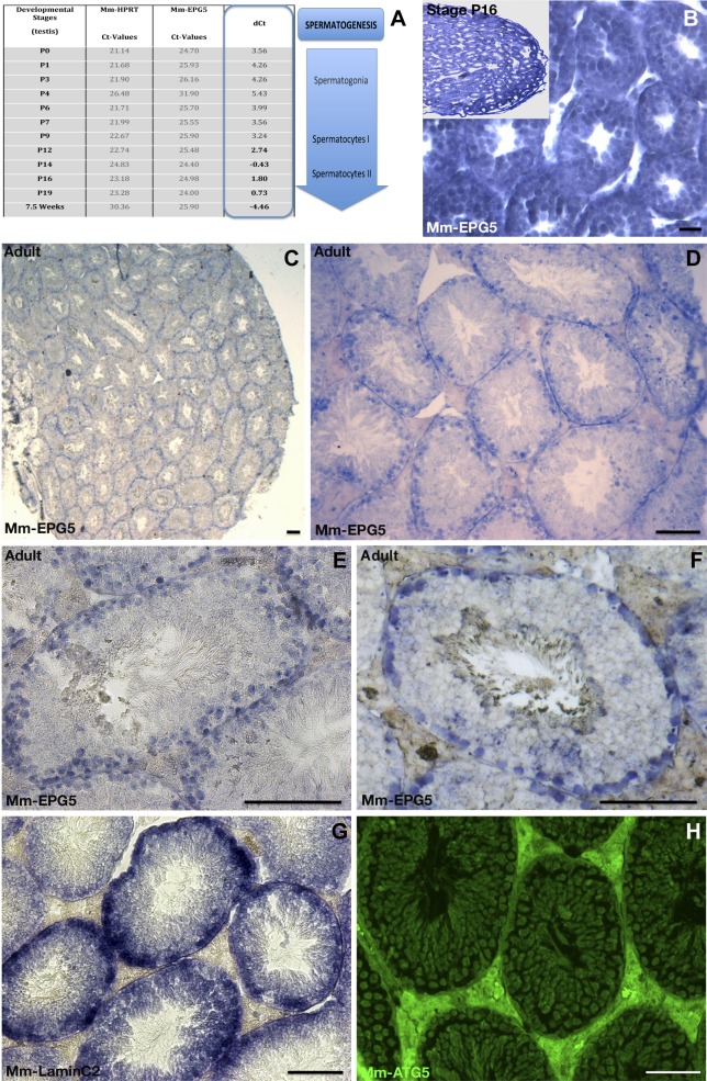
Figure 8.
Testicular expression of Mm-epg5 the mouse homolog of epg5. A) qRT-PCR showed a gradually increasing expression of Mm-epg5 during mouse testicular development from stage P0 up to 7.5 wk. B–F) In situ hybridization of Mm-epg5 at different stages of testis development (blue). B) At stage 16, Mm-epg5 was expressed in spermatogonia/spermatocytes. C–F) In male gonads of adult mice Mm-epg5 expression was restricted to spermatogonia and localized identically to the spermatogonial marker LC2 (F vs. G). F, H) In the adult mouse testis, Mm-epg5 did not colocalize with ATG5. H) In contrast to medaka testis, immunochemistry using an ATG5 antibody revealed that the expression of Mm-ATG5 was not restricted to germ cells, but was more widely and ubiquitously expressed in mouse testis. F vs. H) Mm-epg5 and Mm-ATG5 do not particularly colocalize in spermatogonia cells. Scale bars, 100 µm.

