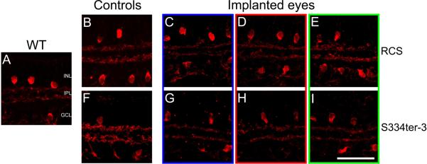
Figure 4.

ChAT labeling in retinal cross-sections from RCS eyes (C-E, implanted from 4 to 8 weeks postnatal) and S334ter-3 eyes (G-I, implanted from 7 to 27 weeks postnatal) with bPVA devices. Both RCS (B-E) and S334ter-3 sections (F-I), like wild-type (A), show typical cholinergic amacrine cell morphology with somata in the INL and GCL and processes in a dual-lamination pattern within the IPL. ChAT labeling patterns in implanted areas (C and G) are identical to those in adjacent (D and H), distal (E and I), Scale bar=50μm.
