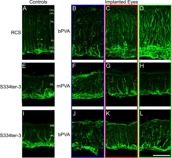
Figure 6.
GFAP labeling in retinal cross-sections from RCS (B-D) with bPVA and S334ter-3 eyes with mPVA (F-H) and bPVA (K-L) devices. RCS were implanted 4 to 8 weeks postnatally, while the S334ter-3 animals were implanted at 12 to 32 weeks. Glial reaction is widespread in all RCS tissue (A-D), but implanted areas (B) do not show increased GFAP labeling in comparison with adjacent (C), distal (D), and age-matched unimplanted control (A) sections. Similar to RCS tissue, S334ter-3 sections (E-L) show widespread gliosis due to photoreceptor degeneration. However, GFAP labeling is not augmented in implanted regions (F and J) relative to adjacent (G and K), distal (H and L), and age-matched unimplanted control section (E). S334ter-3 retinas display a characteristic glial seal above the INL, not seen in wild-type retinas (data not shown), consistent with advanced photoreceptor degeneration. NFL=nerve fiber layer. Scale bar=50μm.

