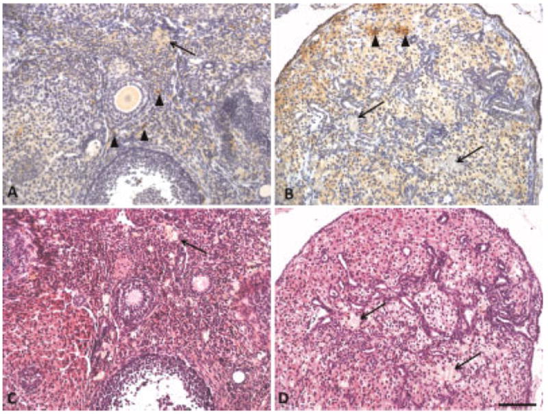
Fig. 6.

F4/80 positive macrophages stained brown in (A) normal ovary and (B) carcinoma. Corresponding H&E sections for (C) normal ovary and (D) carcinoma. Sections also contained foamy cells that are not macrophages. Arrow heads, macrophages; arrow, foamy cells that are not macrophages. Scale bar equals 100 μm.
