Fig. 3.
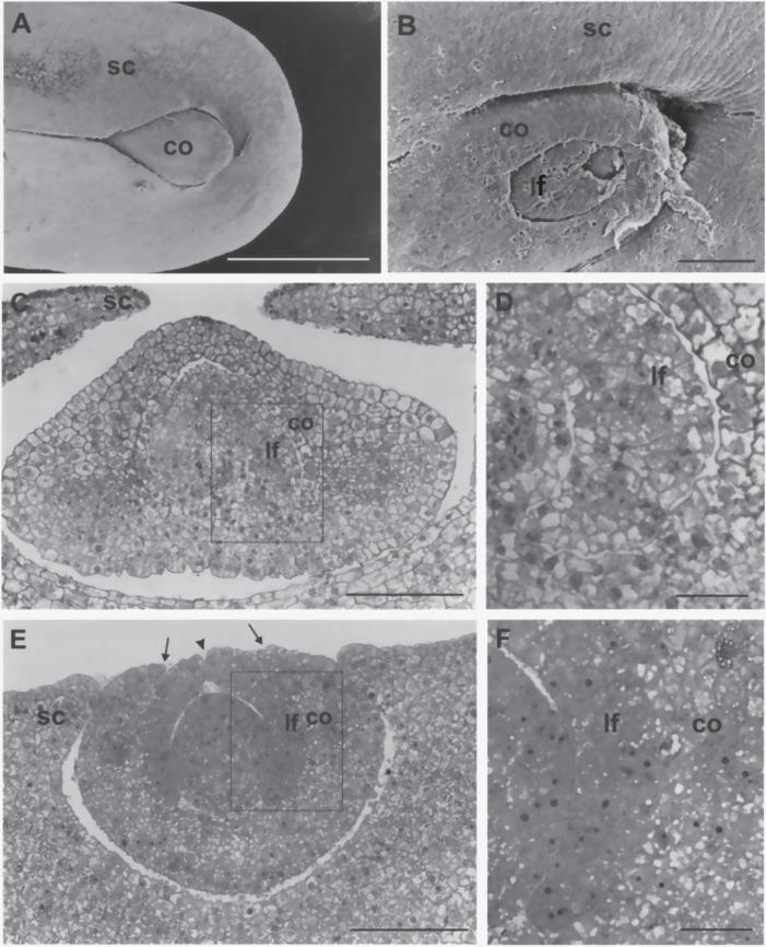
Scanning electron microscope (A, B) and light microscope (C–F) micrographs of the shoot region of 18 DAP embryos. (A) A wild-type embryo with the coleoptile enveloped by the scutellum. (B) A malformed fdl1-1 mutant embryo. Note the less curved scutellum, the uncovered shoot and the openings in coleoptile and first leaf surfaces. (C) The wild-type scutellum encloses the coleoptile, which contains the first leaf. (D) Higher magnification of the area framed in C shows the distinct coleoptile and leaf. (E) Laterally expanded scutellum, open coleoptile surface (arrows) and cleft leaf (arrowhead) are visible in the fdl1-1 mutant embryo shoot. (F) Higher magnification of the area framed in E. Note the lack of tissue identity between coleoptile and leaf in the fused region. co, coleoptile; lf, first leaf; sc, scutellum. Bars: 1mm (A); 100 μm (B, C, E); 25 μM (D, F).

